HOW DO WE PROPERLY DIAGNOSE ORAL CANCER?
Even though all of us is so much in tune to the channel of CoVid-19 pandemic saga, we shouldn't disregard other diseases that are also as deadly or even worse than CoVid-19. That's why DMD Center felt that it is important that we put a spotlight on oral cancer. Why?
1. It is the sixth most common cancer worldwide, with an anticipated yearly incidence of more than 400,000 with the prevalence being higher risk among men. That on average, studies have shown that more than half of those diagnosed with this disease will only survive more than 5 years. The mortality rate for oral cancer is higher in comparison to other form of cancers we routinely hear about, such as cervical cancer, Hodgkin’s lymphoma, laryngeal cancer, testicular cancer, endocrine system, thyroid cancer, or melanoma. Therefore, the condition of probable oral cancer must be taken seriously when we see signs of its existence in our tentative diagnosis from our patient, so, learning how to diagnose it properly is critical in our dental practice.
2. It is also important to take note that the detection of oral cancer occurs in later stage. 65% to 75% of oral cancer lesions are not identified during a visual examination until stages III or IV. Oral cancer 5-year survival is poor, similar to colon-rectal cancer at 62% and significantly lower than for cervical cancer and breast cancer. Those who do survive have not emerged unscathed, having withstood the rigors and painful outcomes of radiation, chemotherapy, and disfiguring surgery. Oral cancer often goes undetected by the patient because in its early stages, it may be symptom-free.
3. Late stage discovery of this disease is due to the fact that the tumor may develop asymptomatic and the patient might recognize only when the cancer has metastasized to another location such as the lymph nodes of the neck. Discovery at this later stage has a negative impact on survival rates due to the metastases as well as deeper invasion of the localized structures. Due to more than half of oral cancers being advanced at the time the cancer is detected, the mortality rate for oral cancer has not decreased in more than 4 decades, according to numerous researches.
4. Oral cancer is usually tobacco-related which is prevalent bad habits nowadays. Disturbing trends in our youth population such as smokeless tobacco products, flavored cigarillos or hookahs (tobacco water pipes)—often accompanied by binge drinking have grown in popularity, and this may present an increased risk for oral cancer at an earlier age. While, if diagnosed in the fifth to seventh decade of life with the added risk factor of age and prolonged, cumulative exposure to tobacco can be indeed deadly to your patient.
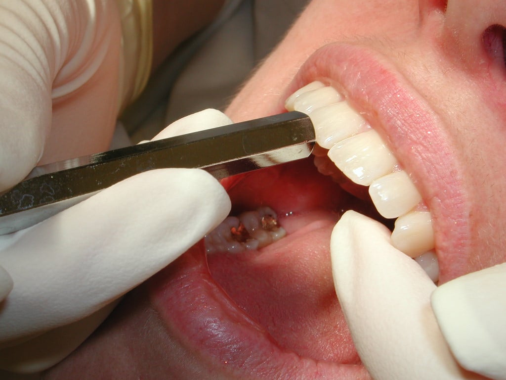
In order to assists you in detecting the need for further oral cancer test and screening, the following is a concise overview of extraoral examination you should do as Part 1 of this post.
SYSTEMIC EXTRAORAL EXAMINATION
STEP 1
To start with the examination, a review and assessment of the systemic health and pharmacological status of the patient is always done prior to any dental examination.
Things that should be checked:
- Last visit to physician
- Last visit to the hospital
- Allergies
- History of systemic or hereditary diseases
- Last medication intake?
- Is there difficulty in swallowing?
- Is there difficulty in breathing?
- Is there difficulty in mastication?
- Is there difficulty in moving your head sideways?
- Is there any presence of bumps or sores inside the mouth?
- Is there difficulty in tasting?
- Are you a smoker?
- How long have you been smoking?
STEP 2
The extraoral examination continues with observation of the head and neck, as well as observation of the sound of the patient's voice and eye movements commencing from when the patient is first seated in the treatment room.
- Hoarseness in the voice may warrant a need further investigation if it has been persistent, since this may be an indication/suspicion of a growth within the larynx/oropharynx.
- Abnormal breathing may be a sign of anxiety or fatigue.
- Pupil size may signify a reaction to drugs or state of emergency as well as an indication of a disease state or inflammatory presence.
- The appearance of the face is further evaluated noting any asymmetry, swelling or discoloration. Inspection of the skin includes the color, texture, the presence of eruptions or swellings, or any abnormal growth.
- Observe all areas of exposed skin, paying particular attention to areas behind the ears and the back of the head and neck.
- Have patients remove their eyeglasses to make certain there are no hidden growths or developments that would have otherwise gone unnoticed. The areas along the hairline and under the eyeglasses will require tactile palpation in order to identify any swellings or abnormal growths.
STEP 3
The next step is examination of the temporomandibular joint. Utilized both hands in examining the temporomandibular joint. This is accomplished by placing your finger pads over the joint just anterior to the ear.
- Instruct the patient to open and close as well as move the jaw to the left and right.
- Check for any limitations or deviations upon opening, subluxation, tenderness, sensitivity.
- Be sensitive to noises such as a grating, clicking, or popping.
STEP 4
The next area to be examined is the parotid salivary glands. The extraoral palpation of the parotid salivary glands is best examined using a bilateral technique, employing light pressure and placing fingers at the angles of the mandible over the parotid glands.
- Compare findings for symmetry for both sides. A normal parotid glands should not be palpable and exhibit no tenderness. An abnormal salivary glands may be painful, swollen or indurated.
STEP 5
The lymph nodes are examined next with the clinician behind the patient and the patient's chin slightly elevated.
- Evaluation is done by a gentle rolling motion of the fingers, using the bilateral palpation technique. Note any enlargement, tenderness, lack of mobility, hardness, or asymmetry.
- If enlargement is detected, the examiner should determine the mobility and consistency of the nodes.
- Enlargement of the lymphnodes or lymphadenopathy may be attributed to either an infectious or inflammatory process or a malignant neoplasm. Clinical characteristics may help discern the difference
STEP 6
Next, submental and submandibular nodes should be examined carefully. With the patient's head back slightly, first examine the submental nodes.
- Instruct the patient to bite together lightly and place the tongue into palatal vault. This results in a tensing of the mylohyoid muscle, allowing for easier palpation of submental glands.
- Moving posterior toward the angle of the mandible and palpating directly below the line of the mandible are the submandibular glands.
STEP 7
Another area to examine are the cervical nodes; both superficial and deep nodes. This set forms a complex chain of numerous nodes.
- Instruct the patient to turn the head in order to reposition the sternocleidomastoid muscle for ease of palpation and better access of both the superficial/deep cervical nodes.
- Note any enlargement, tenderness, lack of mobility, hardness, or asymmetry.
STEP 8
The supraclavicular nodes are palpated next, found superior to the clavicle in the hollow area or supraclavicular fossa directly above the collarbone.
- Note any enlargement, tenderness, lack of mobility, hardness, or asymmetry.
STEP 9
The next nodes to be palpated are the occipital nodes. These are associated with the occipital artery at the posterior base of the skull.
- Using a bilateral technique, palpation is done directly below the base of the occipital bone.
- Reclining the patient's head to the front, exposing the occipital area may facilitate better access for palpation of the occipital nodes.
- Note any enlargement, tenderness, lack of mobility, hardness, or asymmetry.
STEP 10
The posterior auricular and anterior auricular or preauricular nodes. They both in front of the tragus. Both pre and postauricular nodes' efferent vessels drain into the superior deep cervical nodes.
- Check for any signs of enlargement, tenderness, lack of mobility, hardness, or asymmetry.
STEP 11
The thyroid gland, normally not detected by palpation, is examined next.
- An abnormal gland could be indurated, enlarged on one or both sides, or contain palpable nodes. When using bilateral palpation, palpation is done on both sides of the gland, noting any nodules or masse.
- Instruct your patient to swallow, which in turn will elevate the thyroid gland; allowing for an abnormality to become more apparent.
- Asymmetrical movement of the thyroid cartilage during swallowing might indicate that the gland is fixed to underlying tissues.
- If the patient is obese, it may be easier to palpate this area positioned behind the patient, having him or her turn the head toward the examining side.
CONCLUSION
In conclusion, a proper dental examination must be done thoroughly and comprehensively both extraoral and intraoral. If there's a need to delve in further to the oral condition of your patient, a comprehensive oral cancer examination must be done because this serves as the most effective mechanism of protecting your patients' health and by doing so can save your patient's life. It is advisable that with every dental examination you do, including recall appointments, the evaluation of tissues must be included, so, warning signs (abnormalities) of oral cancer or other mucosal pathology can be detected earlier and corresponding treatment can be done immediately.
It is also imperative if certain abnormalities are observed, you should inform the patient to undergo a thorough and comprehensive screening examination, which includes screening for oral cancer. Informing and educating our patients about the risk of this disease can save your patient's life. Failing to advice an oral cancer screening especially when there's an apparent need to do it especially in the presence of an oral lesion can be considered negligence and malpractice on our part.
Contributors:
Dr. Bryan Anduiza – Writer
Dr. Jean Galindez – Writer | Editor
References:
1. Horowitz AM, Drury TF, Goodman HS, et al. Oral pharyngeal cancer prevention and early detection. Dentists' opinions and practices. J Am Dent Assoc. 2000;131:453-462.
2. Hein C, Kunselman B, Frese P. Preliminary findings of consumer-patient's perceptions of dental hygienists' scope of practice/qualifications and the level of care being rendered. American Dental Hygienists' Association Annual Session, Orlando Fla, June 21 to 28, 2006.
3. Bregman JA. Early oral cancer detection: Why you? Why now? oralcancernews.org/wp/early-oral-cancer-detection-why-you-why-now. Accessed on: February 19, 2010.
4. Poh CF, Zhang L, Anderson DW, et al. Fluorescence visualization detection of field alterations in tumor margins of oral cancer patients. Clin Cancer Res. 2006;12:6716-6722.
5. Wong DT. Salivary diagnostics powered by nanotechnologies, proteomics and genomics. J Am Dent Assoc. 2006;137:313-321.
6. American Dental Association. Facts about oral cancer. ada.org/public/topics/cancer_oral.asp#facts. Accessed January 11, 2010.
7. The Oral Cancer Foundation. Oral cancer facts. oralcancerfoundation.org/facts/index.htm. Accessed January 11, 2010.
8. Shah JP, Singh B. Keynote comment: Why the lack of progress for oral cancer? Lancet Oncology. 2006;7:356-357.
9. Petersen PE. Strengthening the prevention of oral cancer: the WHO perspective. Community Dent Oral Epidemiol. 2005;33:397-399.
10. Silverman S, Eversole LR, EL Truelove. Oral premalignancies and squamous cell carcinoma. Essentials of Oral Medicine. Hamilton, Ontario, Canada: BC Decker; 2002:186-187.
11. Ries LAG, Eisner MP, Kosary CL, et al, eds. SEER cancer statistics review, 1973-1998. Bethesda, Md: National Cancer Institute; 2001. seer.cancer.gov/csr/1973_1998/index.html. Accessed January 11, 2010.
12. The Oral Cancer Foundation. The HPV connection. oralcancerfoundation.org/hpv/index.htm. Accessed January 11, 2010.
13. Hammarstedt L, Lindquist D, Dahlstrand H, et al. Human papillomavirus as a risk factor for the increase in incidence of tonsillar cancer. Int J Cancer. 2006;119:2620-2623.
14. Haddad RI. Human papillomavirus infection and oropharyngeal cancer. Medscape CME Course, 2007. cme.medscape.com/viewarticle/559789. Accessed January 11, 2010.
15. MedlinePlus. Oral cancer. nlm.nih.gov/ medlineplus/ency/article/001035.htm. Accessed January 11
