We are often daunted when we have to deal with technical stuff especially when you are born in the era where pen and paper is how you can only write things, when a pager is the only option to send a message and when doing a radiographic examination can only be viewed on a film. We want to stay where we are comfortable with as we feel there’s nothing wrong with it. Why fix it when it ain’t broken, right? Well, I do agree at a certain level. Even I, who loves technology will admit that it still requires serious work from us before one can reap its benefits and when one is clueless about it, I can’t judge you if you rather go back to what works for you even if it is considered outdated. Having said that, please take note that we are not ordinary people, we are health providers and doctors in our own right that deals with the health issues of our patients and most of the time their lives is in our hands, so, we can’t simply ignore what needs to be done for our patients’ sake. Thus, I am writing this blog to let you know that though it does require work, if you put your mind into it, the possibilities of making your diagnosis more accurate and safer to your patients outweigh all the presumed heavy lifting you may do along the way. So, let me put your mind at ease on how the process of understanding 3D digital imaging can be better understood for those who are afraid to take a single step to the age that is digital.
I. Compare It to a Camera. - 3D digital imaging is almost exactly the same as thinking of your camera and when you take photos. It truly has very close similarities because both are used to take images. So, it is not too far out for them to be actually the same in principles. So, when your mind is in a mush on how you will be able to simplify 3D imaging in diagnostics, then, imagine your camera or better yet your phone mounted on a selfie stick. Let’s do simple comparative analysis:

- For your photos the image is comprised of pixels. A pixel is the smallest single component of a digital image. And, 3D digital imaging, it is called a voxel. This is critical because this defines the resolution and the colour depth of your image. The higher the number of pixels in your image, the higher its resolution and quality of your photo. Which means it is the same in voxel.
![]()
- Your camera have different lenses you can attach to it particularly on DSLR (Digital Single-Lens Reflex) while a point and shoot camera has it own settings to provide you lens you may want without the complications that comes along with it as well as the cost. That’s the same we have in 2D digital and 3D digital imaging. In 2D digital imaging, it is like a point and shoot camera. All you got to do is make sure you are on the right direction on how you want to take a photo of your image. Exactly the same principles. While on DSLR, it can be a bit complicated to some and requires that we learn the different lenses, so, we can have better photographs depending on your preferences which is the same principles used in 3D imaging. We need to know where we want to have more focus on the image we need. If the camera have normal, wide angle, telephoto, prime, macro, and zoom lens, the 3D digital imaging has the same principles. You have the focus, medium and large field of view. It is even the same on a selfie, the longer you extend your stick, the bigger the view it can take but the lesser is the focus and resolution is the photo while the shorter you make it, the more focus it becomes on the image you want to take.

- When you are taking a photo, direction or angle of your camera is significant to get the photo you want. It is the same principle on 3D digital imaging. That’s why there’s a Fan Beam CT and a Cone Beam CT scans. You need to be able to identify what you need in order to get the right image you want. And, in Dentistry, a Cone Beam CT Scan is preferred because we need the image to be accurate for our measurements in all angles that are critical in Implant and Endodontics.

II. Each Brand have Its Own Application. - You got to understand that digital camera and digital radiographic imaging equipment are created by different manufacturers and in order for us to be loyal to their brand, they create their own applications. Applications or software allow us to view and manipulate the images we got from their equipment. Thus, once we buy their equipment or use their equipment to take our images, we are required to more or less understand how their applications work. This is more critical in our 3D digital radiographic results. We can have the best digital image result but without understanding how to view it in their application, it is rendered useless and you go back in viewing it on a film which you just wasted either your or your patient’s money of buying it or having it done, respectively.
 III. Required Specifications. - Even at very basic and low level of understanding of how these techy stuff works, I hope you understand that a great image in order to be rendered requires great power. And, great power requires certain specifications on your equipment. Even if you don’t buy a RVG (radiovisiography) periapical x-ray, a digital pano or pano-ceph or a CBCT, you still need a computer with good specifications especially the video card and its RAM to successfully view the images you have requested and acquired from your diagnostic center. This technology will require a minimum standard from your computer for the image/s to render properly and for you to be able to manipulate the image/s without a hassle. You also have to appreciate the fact on how fast technology upgrades every year and this means your computer’s specifications will need an upgrade at least a minimum of every 2 years. If your accountant is truly worth his/her salt in her profession, he/she will tell you that the depreciation value of any computer, phone and most of our techy gadgets is only 2 years. Precisely because technology development is so fast even if we are not. So, it means if you are going to invest on buying the digital imaging equipment, you need to beef up your computer system including having a back-up.
Download here: Carestream Computer System Requirement. Whatever brand you will buy there's similarity to the requirements and this can be used as a guide.
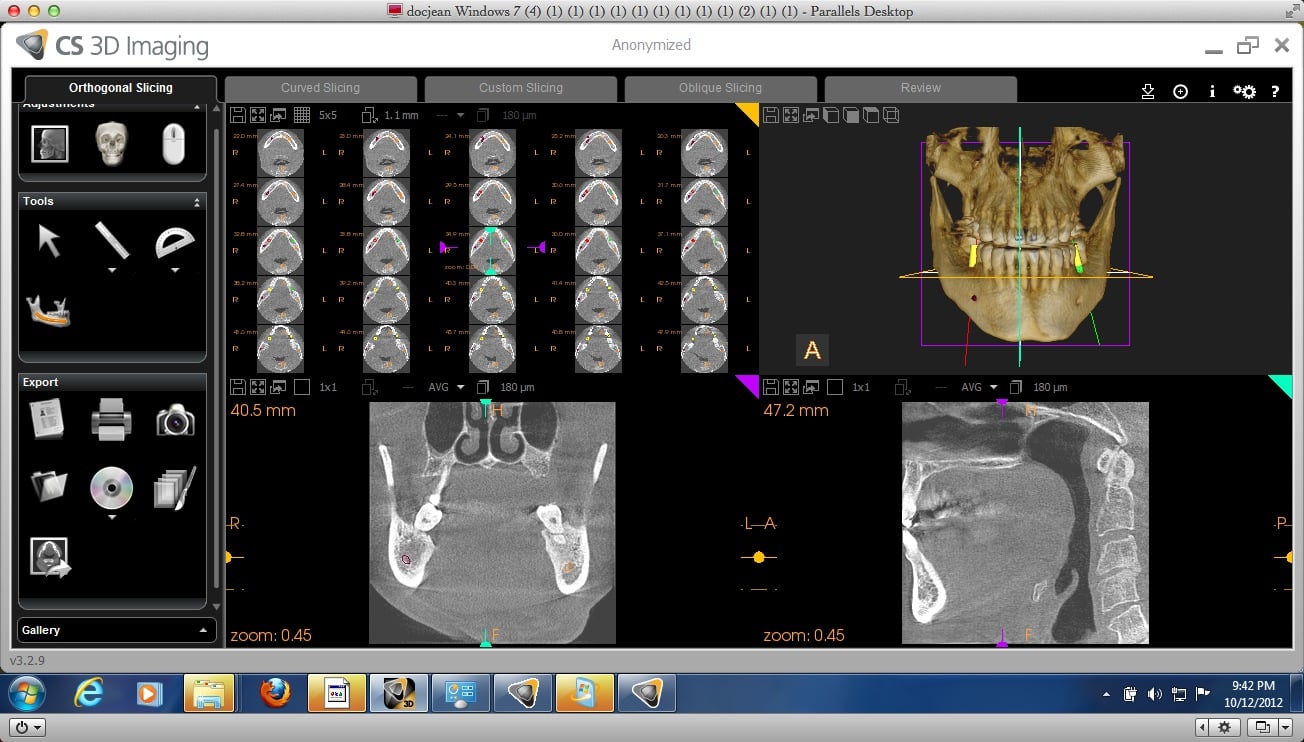
IV. The Slicing. - A 3D digital imaging can be viewed typically on a 4 angles, the Axial, Frontal, Sagittal and MPR (Multi-Planar Reformation). Now depending on your application software you are using, you can actually slice the image according to the level of thickness you want to see. What does it mean on easier terms? It means you have a plank of meat on your chopping board and you need to decide how thick or thin you are going to slice it. The thinner the slice the more you get to see the variations and changes that happens to your meat such as the level of thickness of fat it has. It is the same principle in 3D Digital imaging, the thinner the slice you cut into the image, the more you get to see the change in its anatomy. However, sometimes you don’t need to cut too thin as changes on the anatomy may not occur at a more longer or deeper distance, so, depending on what data you need for your treatment is how you define the thinness and thickness of your slice to the image.
V. The Issue on Radiation Exposure. - There’s a principle in Roentology called ALARA which means “As Low As Reasonably Achievable. It is there for a reason because as much as we want to get an accurate diagnosis to implement an effective treatment, we also need to consider the safety of your patient, your staff and even if you don’t care for them, then, think about yours. We need to follow strictly the safety standards and specifications required when we have radiographic equipment in our clinic. Sometimes we don’t take note on what’s necessary because we are pinching on the money issues and since, you don’t get to actually see what or when it does the greatest damage, it becomes an insignificant consideration to a lot of dental clinics especially in 3rd world countries like ours. As a whole we are already exposed to radiation everyday at a level of 1.8 to 3 millisieverts (mSv). Having said that, it doesn’t mean we go crazy either to the point we totally and extremely go opposite in direction and not do anything. In our profession, visibility is the key and if we don’t see it, we can’t treat it. So, here’s what you need to understand, if you don’t need to see it, then, you don’t need to have it. Just focus only on what you need to see. That’s why there’s a variety of FOV (Field of View) in CBCT. Example, if you are just doing an impaction on the lower left jaw, then, just have that view taken. You don’t need to have the complete scan of the upper and the lower jaw or even the entire lower jaw. Why? Because the bigger image you need to take, the longer the patient will be exposed during the image requisition and the higher the radiation it requires to take that volume of image to render you a quality image result. Plus, not to mention the cost for your patient have to pay. Again, the issue of radiation is not the danger of radiation but the danger of the people dealing with it. As health practitioners we are duty bound to follow what’s good for our patient and to do what’s right base on logical and knowledgeable study of that area. Just remember the greater the quality and field of view of the image required, the higher the radiation is needed to get it. So, don’t get an image if you don’t really need it and make sure your clinic follows to the letter the safety standards or have your patient goes to a diagnostic clinic that does.
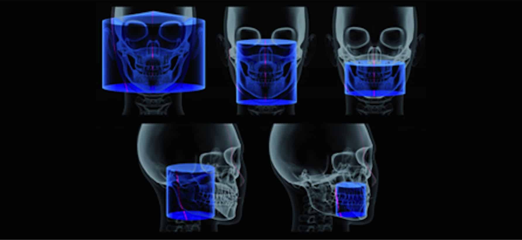 Volume sizes - (a) 15 × 15" (b) 15 × 12" (c) 8 × 10" (d) 8 × 8" (e) 5 × 5" used in cone beam computed tomography imaging
Learning and understanding what we don’t know and what we thought we don’t necessarily need to know before is truly a challenging task. However, it doesn’t mean it can’t be done or you can’t do it. As often as I say or write, I will write it again, it is all base on our attitude and perspective. It is not the technology or the materials out there that is bad that’s why we need to be gangho in changing everything holistic or organic because most of the time, it becomes bad because of the people who use them. We may not like to learn how 3D Digital Imaging works and you may feel you don’t need it because you have done your treatments fairly well so far all these years but it doesn’t mean you can’t make it better or you didn’t actually made a mistake before simply because you don’t know it. The use and utilising the maximum benefits of having better visibility to diagnose and treat your patients for their safety should take precedence on whatever biases we have in learning, understanding and use of 2D and 3D digital imaging. As healthcare providers it is not only the smart thing to do but right thing to do.
[dvk_social_sharing] [et_bloom_inline optin_id="optin_1"]