WHAT ARE THE DIFFERENT SUTURING TECHNIQUES THAT I CAN USE? ITS FEATURES AND BENEFITS.
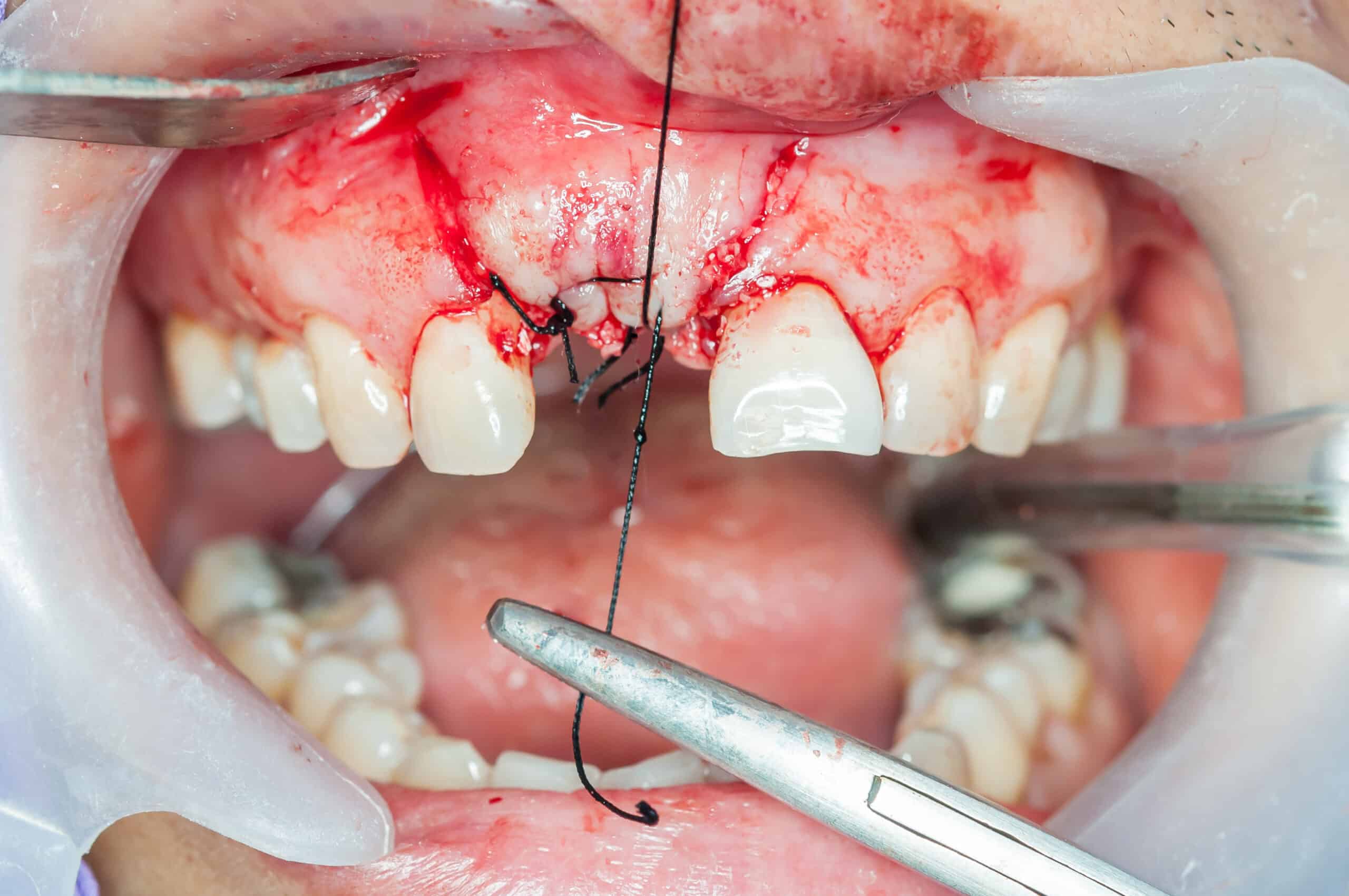
Suturing is important in any dental surgical procedure done on the oral cavity as the choice of technique and material are factors that initiates primary or intentional healing of the surgical wound area. Suturing provides support in coaptation of tissue margins until they heal; can prevent necrosis and possible delay of wound healing; aid in hemostasis and reduces postoperative pain.
It is also best to take note that the selection of materials used such as suturing needles, suture thread and other armamentarium are as important as the suturing technique used in order to achieve success in our treatment. This is because the materials is also a determining the matter in the process of wound healing and keeping the integrity of the soft tissue intact after the procedure. Thus, we will also tackle later on our coming post regarding the types of suturing materials in dentistry.
On this post, though, we will concentrate of familiarizing ourselves about the different types of suturing techniques used in our dental practice, as well as discuss their specific advantages and disadvantages when applied on a surgical procedure.
GENERAL PRINCIPLES IN SUTURING TECHNIQUES
The ability to suture is one of the essential skills required for an oral surgeon or general dentist planning to perform oral surgery. Although, it can appear as just icing in the cake after we are done with the surgery itself, choosing and doing a suturing technique properly requires a sound understanding of the anatomical structure of the tissues, biology of wound healing, good hand–eye coordination, good judgment and finesse. Learning to suture in an expert manner requires an understanding of the proper techniques of needle holding, needle driving, and knot placement. So, here are some general principles in suturing:
1. The tissues should passed through the curve of the needle.
2. The tissue should enter perpendicular to the surface of the needle. Avoid the needle penetrating the tissue obliquely as it may tear the tissues.
3. The needle should be held vertically and longitudinally perpendicular to the needle holder.
4. The needle holder should approximately hold the 1/3 of needle from the end of the curve to avoid slipping while suturing.
5. The needle holder is held by placing the thumb and the fourth finger into the loops and placing the index finger on the fulcrum of the needle holder to provide stability.
6. The distance of the needle passed should be greater than the distance from the tissue edge to avoid tearing of the tissue.
7. The tissue should not be placed under tension / too tightly to avoid necrosis. Undermine the tissue if you see that the suture or tissue is in tension.
8. Each suture should be placed with a distance of 3-4mm. If the suture is placed closer than 3mm, it should be place in areas of tension to help holding the suture place while movement.
9. If one tissue is side is thinner than the other, the needle should passed from the thinner tissue to the thicker one.
10. If one tissue is side is deeper than the other, the needle should passed from the deeper tissue to the superficial side.
11. The edges of the tissue should be everted and tissue merely approximated with the suture.
12. The knot shpuld be positioned on top of the incision line.
SUTURING TECHNIQUES USED IN DENTISTRY
I. Vertical Mattress Suture
Also called as Internal Vertical Mattress, this suture gives maximum tissue adaptation of the wound site decreasing the amount of dead tissue/ space along the injury line. Used for closing deep wounds.
The needle is passed from one edge to the other and again from the latter edge to the fist and the knot is tied close to it. When needle is brought back from second flap to the first, depth of penetration is more superficial.
The width of the stitch should be increased in proportion to the amount of tension on the wound—that is, the higher the tension, the wider the stitch.
 Modified Vertical Mattress Suture
 Internal Vertical Mattress Suture
 External Vertical Mattress Suture
Advantages of Vertical Mattress Suture
1. Does not interfere with the healing as suture runs parallel to the blood supply.
2. It reduces the amount of dead space during wound healing and provides increased strength across the wound.
Disadvantages of Vertical Mattress Suture
1. Fine wound edges are difficult to approximate using suturing techniques leading to open edges.
2. Sutures need to be removed early to prevents prominent suture marks on the healing soft tissue surfaces.
II. Simple Interrupted Suture
Most commonly used. Inserted singly through side of the wound and tied with a surgeon’s knot. Most common knot used in dental procedures such as extraction, impaction, etc.
Advantage of Simple Interrupted Suture technique
1. Can be used in areas where tissues are under stress.
2. The degree of eversion is produced.
3. Suture are placed 4-8mm apart and can be used to close large wounds where tension is shared between each one separately.
4. In case of loosening of one suture other won’t be affected.
5. In case of any secondary infection or hematoma, suture can be removed while other suture can be left in place.
6. Easy to clean between sutures as there are no interference and not connected.
III. Simple Continuous or Running Suture
This type of suture is placed with the needle inserted in a continuous fashion from one end of the soft tissue to the other such that the suture passes perpendicular to the incision line belowe and obliquely above.
Advantages of Simple Continuous or Running Suture
1. Rapid technique and distributes tension uniformly.
2. More water tight closure.
3. Only 2 knots with associated tags.
Disadvantage of Simple Continuous or Running Suture
If cut at one point, suture slackens along the whole length of the wound, which will then gape open.
IV. Continuous Locking Suture
This technique is indicated on long edentulous areas along with the retromolar or tuberosity areas. This type of suture is similar to continuous suture but the locking is done by inserting the suture through its own loop while closing the suture.
Advantages of Continuous Locking Suture
1. Watertight closure, cannot be used in areas of tension.
2. Avoid multiple knots.
3. Prevents excessive tightening.
4. Distributes tension uniformly.
Disadvantages of Continuous Locking Suture
Prevents adjustment of tension over suture line as tissue swelling occurs.
V. Horizontal Mattress Suture
This suturing techniques everts mucosal margins, bringing greater areas of raw tissue into contact. So used for closing bony deficiencies such as oro-antral fistula or cystic cavities.
To place a horizontal mattress suture, the needle has to pass through one edge of the incision to the other and it is again brought back and inserted into the first edge. This process is continued along with the incision line to the other end edge of the incision and a knot is given.
Advantages of Horizontal Mattress Suture
1. Prevents the flap from being inverted into the cavity.
2. Use to control post-operative hemorrhage from gingiva around the tooth socket to tense the mucoperiosteum over the underlying bone.
3. It does not cut through the tissue, so used in case of tissue under tension (inadequate tissue).
Disadvantages of Horizontal Mattress Suture
1. Difficulty in insertion.
2. Constricts the blood supply to the incision if improperly used, cause wound necrosis and dehiscence.
VI. Figure of 8 or Criss-Cross Suture
This suture is used over edentulous spaces. When beginning this technique, a 3/8 circle needle penetrates at the level of the mucogingival junction at the mesiobuccal line, travels horizontally under the flap, and emerges at the distobuccal line angle, the procedure is done on the lingual aspect,the suture material crosses over the surgical field, tying of suture knot on buccal aspect forming a cross on the flap.
Advantages of Figure of 8
1. It is removable. Traditional subcutaneous sutures cannot be removed and must be resorbeable. In other words, it must be made of material that can be absorbed by the body’s tissue enzymes.
2. It allows closure of two layers simultaneously.
3. Compared with the interrupted stitch, ischemia at the edge of the suture is reduced. Ischemia is a reduction in the blood supply to the body tissue.
4. Being removable, one avoids burying foreign material in the depth of the tissue, minimizing the chances of developing a stitch abscess or related complications.
5. This technique enables any length difference between the flaps to be evened up when sutured.
6. It minimizes “dog’s ear” defects.
Disadvantages of Figure of 8
1. The figure-8 suturing technique is more difficult to master and perform properly compared to interrupted sutures.
2. Patients experience slightly more discomfort on stitch removal compared to the interrupted sutures.
VII. Sling Suture Techniques
The sling suturing technique is the technique of choice when the goal of therapy is to reposition one of the surgical flaps at a particular occlusal apical level that is independent of the other gingival tissue height. The sling suture is widely used for root coverage, gingiva esthetics, open flap implant surgery and etc.
The suture could be applied both buccal/ lingual side. The 3/8 circle reverse cutting needle is first passed under the distal contact point of the most distal interdental papilla then the suture needle pierces through the inner side of the elevated surgical flap 3mm from the tip of the papilla, passage of the suture needle back under the contact point, then passed under the next contact point in a mesial direction and then the needle pierces through the inner surface of the elevated surgical flap 3mm from the tip of the interdental papilla, then passage of the needle back under the contact point, tying of the suture knot on the non elevated tissues.
Advantages of Sling Suture Techniques
Elimination of the need to place additional sutures.
CONCLUSION
As dentists knowing the proper technique of suturing and when to apply them is very important as this enhances the approximation of the wound edges, which helps minimize and redistribute the tension in the tissues. Moreover, knowing which suturing technique to be used on your case and implementing them correctly also helps maintain the level of hemostasis in which the tissue needs during an operation as such optimize its healing process afterwards.
CONTRIBUTORS:
Dr. Bryan Anduiza - Writer
Dr. Mary Jean Villanueva - Editor
REFERENCES:
1. Edward S Cohen Atlas of cosmetic and reconstructive periodontal surgery 2nd Edn
2. Harry Dym Atlas of Minor Oral Surgery.
3. Baldi C, Pini Prato G, Pagliaro U, Nieri M, Saletta D, et al (1999) Coronally advanced flap procedure for root coverage. Is flap thickness a relevant predictor to achieve root coverage? A 19-case series. J Periodontol 70(9): 1077-1084.
4. Braun, Aesculap (2006) Suture Glossary.
5. Chrimax (2001) Non-absrobable Materials: Reactionin Tissue
6. Dunn DL (2007) Wound Closure Manual. Johnson and Johnson Engineering. Toolbox (2012) Stiffness. (2012) Engineering Toolbox, Stiffness.
7. (1985) Wound closure manual, Ethicon, Somerville, New Jersey, USA.
8. Helmenstine AM (2012) Strain About.com Chemistry
9. Goeffrey L.Howe, Minor Oral Surgery.
10. Najibi S, Banglmeier R, Matta JM, Tannast M (2010) Material Properties of Common Suture Materials in Orthopaedic Surgery. Iowa Orthopaedic Journal 30: 84-88.
11. W Harry Archer, Oral and Maxillofacial Surgery
12.(2004) Oral tissue reaction to suture materials: A review Periodontal Abstracts: 52: 237-244.
13. Postlethwait RW, Willigan D A, Ulin AW (1975) Human Tissue Reaction to Sutures. Annals of Surgery 181(2): 144-150.
14. Ratner BD, Hoffman A, Schoen F, J Lemons (2004) Surface Properties and Surface Characterization of Materials. Biomaterial Science: An Introduction to Material in Medicine. (2nd edn). San Diego, US.
15. Salhan S, Dass A (2012) Textbook of Gynecology. Jaypee Brothers Medical, New Delhi, India.
16. Surgical Knot tying Ethicon manual, Somerville, New Jersey, USA.
17. Suture material techniques and knots. Serag wieesner, Naila, Germany.
18. Sandro Siervo, Suturing techniques in oral surgery.
19. Carranza , Textbook of clinical periodontology, 10th edn
20. https://www.juniordentist.com/suturing-techniques-used-in-dentistry.html
21. Hassan H Koshak (2017), Dental Suturing Materials and Techniques
