HOW DO WE PROPERLY DIAGNOSE ORAL CANCER?
NOTE: This is the Part 2 of Addressing Oral Cancer. Please read Part 1 first of the Post, Click this: PART 1
This is the continuation of Part 1 and let's all dive into more information to guide you all further.
SYSTEMIC INTRAORAL EXAMINATION
It is best to follow a systematic and consistent approach when performing the intraoral examination. The following is a suggested 7-step systematic approach:
STEP 1
- The lips should have a normal/well-defined vermilion border and be even in coloration.
- Use the method of bidigital palpation to note any swelling, induration or observed texture or color change.
- Take note of dryness and/or unclear demarcation of lip vermillion and skin exist should be noted as "lip at risk" to flag the area for subsequent examinations. Also examine for loss of vertical dimension manifested often on labial commissures with the outcome being angular cheilitis.
- Further investigation to determine the causative factor behind the loss of vertical dimension would be warranted. Reinforce the need for sunblock protection, especially related to those patients who are active outdoors and have prolonged exposure to sunlight. Sunblock protection for the lips has had a positive effect on reducing the number of cancers related to the lip.
STEP 2
Inspect the labial mucosa.
- Inspect through a visual and tactile method.
- Instruct the patient to partially open the mouth, allowing examination of the labial mucosa and sulcus of the maxillary and mandibular vestibule and frenum.
STEP 3
Inspect the buccal mucosa using visual inspection and tactile palpation.
- This is best accomplished by using a bidigital palpation technique with the thumb placed against the buccal mucosa simultaneously with external palpation.
- Note for any change in pigmentation, texture or diminished mobility or other abnormalities of the mucosa.
- Inspect the parotid gland from the intraoral aspect at this time as well as palpating both the maxillary tuberosities and retromolar pads.
STEP 4
Examine the gingival tissues.
- Observe attached and free gingiva on both arches, assessing for normal color and contour using digital palpation.
- Use a 2-x-2 gauze to dry the tissues to provide an enhanced assessment.
STEP 5
Inspect all surfaces of the tongue.
- The tongue is a very high-risk area for oral cancer as well as for candida infections. Candida infections can be an indication of an underlying systemic disease.
- The tongue should be examined thoroughly using both visual and tactile methods.
- Visual inspection alone is inadequate in its ability to identify early changes to the mucosal surface of the tongue.
- It is best to follow a systematic approach when inspecting the tongue, commencing with examination of the dorsum, then lateral borders and concluding with the ventral surface. The dorsum is the first area of the tongue to be examined.
- Ask the patient to protrude the tongue, moving from side to side, noting any abnormality of mobility or restriction of movement.
With the patient's tongue at rest, and mouth partially open, inspect and palpate the dorsum of the tongue to detect any swelling or fixed mass.
The next to examine is the lateral borders of the tongue. Usually, the common site for oral cancer is on this lateral aspect of the tongue. - Retract the cheek, inspect the left and right lateral margins of the tongue.
- Use a piece of gauze to handle the tip of the tongue to further protrude the tongue. This will aid in the visualization of the most posterior aspects of the tongue's lateral borders, including the lingual tonsils.
- With the tongue fully protruded (held and manipulated forward and side by side by the clinician for optimal visual access), inspect the posterior aspect and base of tongue using digital palpation along the lateral borders to identify any changes in tissue texture or consistency, noting any swelling/induration. If detected, compare with the opposing lateral border. Be suspicious of an abnormality that is unilateral. The last area of the tongue to be examined is the ventral surface. Instruct the patient to touch the roof of the mouth with the tip of the tongue.
- This will allow full inspection of the ventral surface of the tongue. Palpate the ventral surface of the tongue to aid in any detection of growths, swelling or area of tenderness, as well as any color or texture changes.
STEP 6
Examine the floor of the mouth carefully, keeping in mind that this is another highly vulnerable area that requires close and thorough inspection. Areas are easily hidden from visual inspection.
- Instruct the patient to touch the roof of the mouth with the tip of the tongue.
- Inspect the floor of the mouth for changes in color, texture, swellings, or other surface abnormalities.
- Using bimanual palpation, compress the floor of mouth against the opposite hand. This is the only effective way to identify any area of firmness or mass as well as locating any feeling of tenderness.
STEP 7
Inspection of the oropharynx and palatal tissues.
- Check the entire area of the oropharynx, examining the tonsil region including the uvula, tonsillar pillars, and palatine tonsils for presence, color, size, or any noted abnormalities.
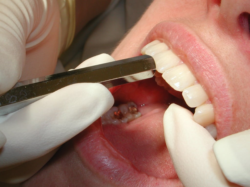
- When examining the oropharynx, it is best to depress the tongue down toward the floor of the mouth using either a tongue blade or the back of the mouth mirror while instructing the patient to take a deep breath and hold or say "ah".
- This method enables the clinician to gain better visual access of the oropharynx area. The soft palate should be visually examined next, accompanied by digital palpation of the hard palate, noting any asymmetries, swelling or mucosal changes.
CONCLUSION
In conclusion, a proper dental examination must be done thoroughly and comprehensively both extraoral and intraoral. If there's a need to delve in further to the oral condition of your patient, a comprehensive oral cancer examination must be done because this serves as the most effective mechanism of protecting your patients' health and by doing so can save your patient's life. It is advisable that with every dental examination you do, including recall appointments, the evaluation of tissues must be included, so, warning signs (abnormalities) of oral cancer or other mucosal pathology can be detected earlier and corresponding treatment can be done immediately.
It is also imperative if certain abnormalities are observed, you should inform the patient to undergo a thorough and comprehensive screening examination, which includes screening for oral cancer. Informing and educating our patients about the risk of this disease can save your patient's life. Failing to advice an oral cancer screening especially when there's an apparent need to do it especially in the presence of an oral lesion can be considered negligence and malpractice on our part.
Contributors:
Dr. Bryan Anduiza – Writer
Dr. Jean Galindez – Writer | Editor
References:
1. Horowitz AM, Drury TF, Goodman HS, et al. Oral pharyngeal cancer prevention and early detection. Dentists' opinions and practices. J Am Dent Assoc. 2000;131:453-462.
2. Hein C, Kunselman B, Frese P. Preliminary findings of consumer-patient's perceptions of dental hygienists' scope of practice/qualifications and the level of care being rendered. American Dental Hygienists' Association Annual Session, Orlando Fla, June 21 to 28, 2006.
3. Bregman JA. Early oral cancer detection: Why you? Why now? oralcancernews.org/wp/early-oral-cancer-detection-why-you-why-now. Accessed on: February 19, 2010.
4. Poh CF, Zhang L, Anderson DW, et al. Fluorescence visualization detection of field alterations in tumor margins of oral cancer patients. Clin Cancer Res. 2006;12:6716-6722.
5. Wong DT. Salivary diagnostics powered by nanotechnologies, proteomics and genomics. J Am Dent Assoc. 2006;137:313-321.
6. American Dental Association. Facts about oral cancer. ada.org/public/topics/cancer_oral.asp#facts. Accessed January 11, 2010.
7. The Oral Cancer Foundation. Oral cancer facts. oralcancerfoundation.org/facts/index.htm. Accessed January 11, 2010.
8. Shah JP, Singh B. Keynote comment: Why the lack of progress for oral cancer? Lancet Oncology. 2006;7:356-357.
9. Petersen PE. Strengthening the prevention of oral cancer: the WHO perspective. Community Dent Oral Epidemiol. 2005;33:397-399.
10. Silverman S, Eversole LR, EL Truelove. Oral premalignancies and squamous cell carcinoma. Essentials of Oral Medicine. Hamilton, Ontario, Canada: BC Decker; 2002:186-187.
11. Ries LAG, Eisner MP, Kosary CL, et al, eds. SEER cancer statistics review, 1973-1998. Bethesda, Md: National Cancer Institute; 2001. seer.cancer.gov/csr/1973_1998/index.html. Accessed January 11, 2010.
12. The Oral Cancer Foundation. The HPV connection. oralcancerfoundation.org/hpv/index.htm. Accessed January 11, 2010.
13. Hammarstedt L, Lindquist D, Dahlstrand H, et al. Human papillomavirus as a risk factor for the increase in incidence of tonsillar cancer. Int J Cancer. 2006;119:2620-2623.
14. Haddad RI. Human papillomavirus infection and oropharyngeal cancer. Medscape CME Course, 2007. cme.medscape.com/viewarticle/559789. Accessed January 11, 2010.
15. MedlinePlus. Oral cancer. nlm.nih.gov/ medlineplus/ency/article/001035.htm. Accessed January 11
