Your Know How On 3D Imaging
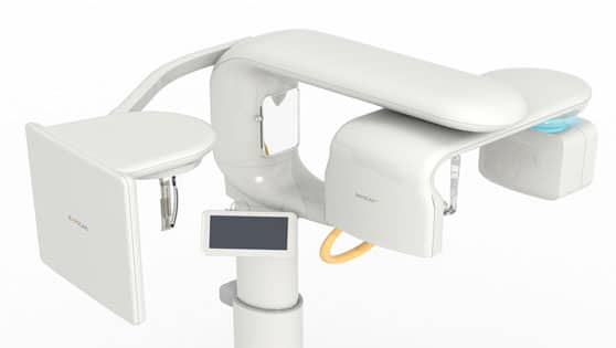
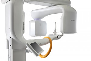
A Cone Beam CT (Computed Tomography) is the standard of care for imaging for placement of dental implants. A traditional CT performed in a hospital has around 80% more radiation than a Cone Beam CT. A Cone Beam CT scanner uses a cone shaped x-ray beam rather than a conventional linear fan beam, as is the case with Medical CT, to provide images of the bony structures of the skull. As result, the medical CT scanner provides a set of consecutive slices of the patient while the Cone Beam CT scanner provides a volume of data.
CBCT scanners are based on volumetric tomography, using a 2D extended digital array providing an area detector. This is combined with a 3D x-ray beam. The cone-beam technique involves a single 360° scan in which the x-ray source and a reciprocating area detector synchronously move around the patient’s head, which is stabilized with a head holder. At certain degree intervals, single projection images, known as “basis” images, are acquired. These are similar to lateral cephalometric radiographic images, each slightly offset from one another. This series of basis projection images is referred to as the projection data. Software programs incorporating sophisticated algorithms including back-filtered projection are applied to these image data to generate a 3D volumetric data set, which can be used to provide primary reconstruction images in 3 orthogonal planes (axial, sagittal and coronal). Although the CBCT principle has been in use for almost 2 decades, only recently — with the development of inexpensive x-ray tubes, high-quality detector systems and powerful personal computers — have affordable systems become commercially available.
- X-ray Beam Limitation Reducing Radiation
- Image Accuracy
- Rapid Scan
- TimeDose Reduction
- Display Modes Unique to Maxillofacial Imaging
- Reduced Image Artifact
[dvk_social_sharing] [et_bloom_inline optin_id="optin_1"]
